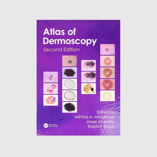
An Atlas Of Dermoscopy, Second Edition
ISBN: 9780415458955
Autores: Marghoob Ashfaq A, Malvehy Josep, Braun Ralph P
Building on a successful first edition, this revised and extended Atlas of Dermoscopy demonstrates the state of the art of how to use dermoscopy to detect and diagnose lesions of the skin, with a special emphasis on malignant skin tumours.
- 9780415458955
- Dermatología
Building on a successful first edition, this revised and extended Atlas of Dermoscopy demonstrates the state of the art of how to use dermoscopy to detect and diagnose lesions of the skin, with a special emphasis on malignant skin tumours. With well over 1,500 photographs, drawings, and tables, the book has extensive clinical correlation with dermoscopic images, so readers can appreciate the added benefits of dermoscopy by comparing the clinical morphology seen with the naked eye with the corresponding dermoscopic morphology; extensive illustrations from the image collections of internationally recognized experts, who have years of experience refining their techniques; and extensive schematic drawings to help readers single out the key structures and patterns to recognize in the dermoscopic images. The second edition has important new material on such topics as observed differences between polarized and non-polarized dermoscopy, newly recognized structures and patterns, refined and revised suggestions for pattern analysis, dermoscopy of the hair and nails, and how to integrate dermoscopy into general clinical practice. It also covers dermoscopically equivocal, false negative, and false positive lesions; discusses further indications for dermoscopy beyond skin cancer; and details common checklists of criteria and algorithms used to diagnose skin lesions
Tabla de contenido:
Introduction Ashfaq A. Marghoob, Ralph P. Braun, and Josep Malvehy Principles of dermoscopy and dermoscopic equipment Steven Q. Wang, Ashfaq A. Marghoob, and Alon Scope Histopathologic tissue correlations of dermoscopic structures Ralph P. Braun, Alon Scope, Ashfaq A. Marghoob, Katrin Kerl, Harold S. Rabinovitz, and Josep Malvehy Two-step algorithm: Differentiating melanocytic from nonmelanocytic lesions Ashfaq A. Marghoob and Ralph P. Braun Nonmelanocytic lesions Basal cell carcinoma Steven Q. Wang and Scott W. Menzies Actinic keratosis, Bowen's disease, keratoacanthoma, and squamous cell carcinoma Iris Zalaudek, Alan Cameron, and Cliff Rosendahl Solar lentigines, seborrheic keratoses, and lichen planus-like keratosis Steven Q. Wang, Harold S. Rabinovitz, Margaret C. Oliveiro, and Ashfaq A. Marghoob Vascular lesions Pedro Zaballos, Josep Malvehy, and Susana Puig Dermatofibromas Pedro Zaballos, Susana Puig, and Josep Malvehy Step 2: Methods to differentiate between melanoma and benign melanocytic nevi Analytic features of melanoma vs benign nevi on nonglabrous skin: Colors and structures Josep Malvehy and Susana Puig Pattern analysis Ashfaq A. Marghoob ABCD rule Ulrike Weigert, Walter H.C. Burgdorf, and Wilhelm Stolz Menzies method Scott W. Menzies Seven-point checklist and the seven rules not to miss melanoma incognito Giuseppe Argenziano CASH algorithm Jennifer A. Stein and Alfred W. Kopf Three-point checklist Giuseppe Argenziano Revised version of pattern analysis Harald Kittler, Cliff Rosendahl, and Alan Cameron Benign melanocytic lesions Congenital melanocytic nevi Ashfaq A. Marghoob Dysplastic (atypical) nevi Ashfaq A. Marghoob and Alfred W. Kopf Banal nevi and growing nevi Iris Zalaudek Blue nevi and variants Cristina Carrera, Josep Malvehy, and Susana Puig Spitz and Reed nevi Gerardo Ferrara and Giuseppe Argenziano Recurrent (persistent) nevi Ashfaq A. Marghoob Melanoma Superficial spreading melanoma Scott W. Menzies Acrolentiginous melanoma Josep Malvehy and Susana Puig Nodular melanoma Scott W. Menzies Lentigo maligna melanoma Alon Scope, Steven Q. Wang, and Harold S. Rabinovit Amelanotic and hypomelanotic melanoma Juergen F. Kreusch, Scott W. Menzies, and Giuseppe Argenziano Equivocal lesions, false positive, and false negative Susana Puig, Josep Malvehy, Ashfaq A. Marghoob, and Giuseppe Argenziano Desmoplastic melanoma and metastatic melanoma Steven Q. Wang, Natalia Jaimes-Lopez, and Ashfaq A. Marghoob Special locations Palms and soles Josep Malvehy and Susana Puig Nails Ralph P. Braun and Luc Thomas Face Nancy H. Kim, Margaret C. Oliviero, Ralph P. Braun, Ashfaq A. Marghoob, and Harold S. Rabinovitz Hair and scalp (trichoscopy) Ralph M. Trueb, Lidia Rudnicka, Malgorzata A. Olszewska, Ralph P. Braun, Isabel Kolm, and Adriana Rakowska Mucosal lesions Andreas Blum and Olga Simionescu
Product Details
Ficha técnica
- Año
- 2012
- Editorial
- Taylor & Francis Ltd
- Páginas
- 384
- Alto
- 28 cm
- Ancho
- 22 cm
- Edición
- 2


